01 — Technology Approach
From "Pulse Signals" to
"Scanning Imaging"
Traditional Flow vs. LASE Technology Approach
Traditional Flow Cytometers:
-
Large bar-shaped laser excitation (e.g., 10μm × 80μm)
-
Records the integrated pulse signal of an entire cell
The LASE Technology proposes a wholly new approach: constructing a linear array of laser spots to perform point-by-point spatial scanning of high-speed flowing cells, and encodes cell images into the detector's time-domain signals.
This means:
-
Cells still flow at several meters per second
-
Clear 2D images are obtained for every detection channel
-
While maintaining high-throughput detection capability
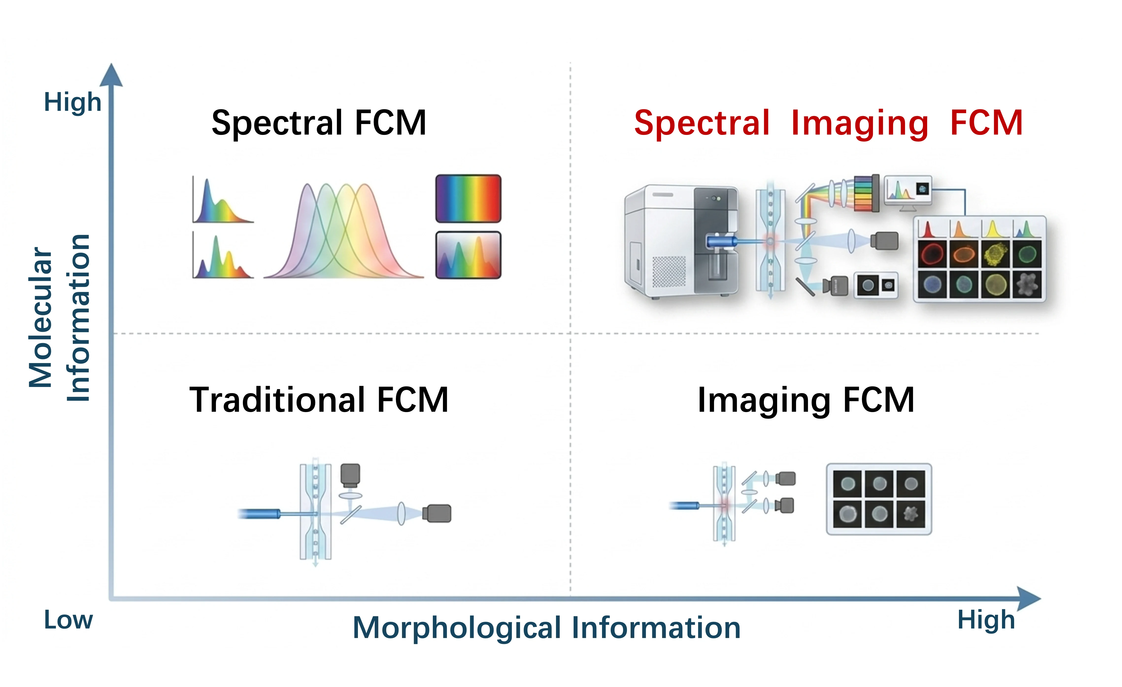
Compatible with both label-free and fluorescence imaging, combined with spectral detection modules, the LASE Technology enables unprecedented spectral imaging detection, with every spectral channel capable of imaging. The imaging process is similar to Laser Scanning Confocal Microscopy. It requires no complex "computational imaging" procedure.

Imaging flow cytometry using linear array spot excitation. Device 1, no. 6 (2023).